
Our Office
Visit Us Online


At the office of Maryville Family Dentistry, we view oral exams as the foundation of long-term dental health. Regular, careful evaluations allow our team to identify small changes early, tailor preventative strategies, and keep your smile functioning comfortably for years. An oral exam is more than a quick look — it’s a focused review of your teeth, gums, bite, and the surrounding tissues that together paint a clear picture of your current oral health.
Your initial oral exam is designed to create a clear starting point for ongoing care. We begin with a thorough review of your medical and dental history so we understand any medications, past treatments, or health conditions that could affect your mouth. This conversation helps us prioritize what to examine and shapes any recommended diagnostic imaging for a complete picture.
During the clinical portion of the exam, we inspect each tooth, the gum tissues, and the supporting bone for signs of wear, decay, inflammation, or abnormal lesions. We also evaluate your bite and jaw joint function to spot patterns of clenching, grinding, or uneven forces that can cause long-term damage. Where appropriate, we perform an oral cancer screening and review any symptoms you may have noticed in the mouth, head, or neck.
Diagnostic images — such as bitewings or periapical films — are taken when they provide useful information beyond what we can see visually. These images reveal hidden decay, check root and bone health, and support accurate treatment planning. When the exam is complete, we’ll summarize our findings, answer your questions, and outline the next steps so you leave with a clear, practical plan for care.

Oral health and general health are closely connected. Inflammation or infection in the mouth can affect other systems in the body, and conversely, systemic conditions often show early signs inside the mouth. During an oral exam, we look for clues such as unexplained gum bleeding, persistent dry mouth, oral lesions, or rapid changes in oral tissues that might warrant coordination with your medical provider.
Research continues to highlight links between chronic oral inflammation and conditions such as cardiovascular disease, diabetes, and respiratory problems. Identifying and controlling oral disease not only preserves teeth and gums, it can also reduce a source of chronic inflammation that may influence your broader health. That makes routine exams an important part of an overall wellness strategy.
For patients with existing medical conditions, we tailor our approach to minimize risk and improve outcomes. Simple adjustments in scheduling, hygiene recommendations, or collaboration with your physician can make dental care safer and more effective. Our goal is to treat oral findings in a way that supports your whole-body health, not just the mouth alone.
Regular checkups and professional cleanings are the most reliable way to prevent small problems from growing into larger ones. Plaque and tartar build up over time in areas that can be difficult to reach with home brushing and flossing alone. During a cleaning, our hygienist removes this buildup, polishes the teeth, and leaves your mouth feeling fresh — while reducing the bacteria responsible for cavities and gum disease.
Beyond polishing and scaling, routine visits are an opportunity for hands-on education. We demonstrate effective brushing and flossing techniques, review tools that suit your needs, and recommend daily habits that protect your smile. For children, these visits are also a chance to monitor development and provide age-appropriate guidance that encourages lifelong healthy habits.
We generally recommend scheduling exams and cleanings at intervals that match each patient’s risk profile. Some people benefit from twice-yearly care, while others may need more frequent monitoring. Whatever the interval, consistent preventative care helps us detect early changes — when treatments tend to be simpler, faster, and less invasive.
Visual exams are essential, but radiographs (x-rays) and other imaging tools reveal what cannot be seen with the naked eye. These diagnostic images help us detect interproximal decay between teeth, check the integrity of tooth roots, assess bone levels around teeth, and identify issues such as impacted or developing teeth. When used appropriately, imaging is a highly informative part of a comprehensive exam.
Digital radiography has become the standard because it produces high-quality images quickly with reduced radiation exposure compared to older film methods. Digital images can be enhanced, measured, and securely stored in your chart for easy comparison over time. They also support more precise treatment planning, whether we’re evaluating a root, planning a restoration, or monitoring healing after treatment.
In certain situations — for example, complex extractions, implant planning, or when detailed anatomic information is required — three-dimensional imaging such as cone-beam computed tomography (CBCT) provides unparalleled detail. CBCT scans allow us to view teeth, bone, and surrounding structures in three dimensions, improving diagnostic accuracy and helping to guide safe, predictable care.

Different imaging tools serve different diagnostic purposes. Small intraoral films like periapicals show an entire tooth from crown to root and are useful for assessing root health and periapical conditions. Bitewing images focus on the crowns of molars and premolars and are particularly good at revealing early decay between teeth. A full mouth series provides a comprehensive set of intraoral views when a complete assessment is needed.
Extraoral options offer broader perspectives. A panoramic film gives a wide view of both jaws and all teeth, helping us evaluate tooth development, impactions, and jawbone form. Cephalometric images are specialized frontal or profile views used primarily in orthodontic assessment. When three-dimensional anatomy is required, CBCT captures volumetric images that support precise diagnosis and surgical planning.
We recommend imaging selectively, guided by clinical findings and your individual needs. Our emphasis is always on using the right tool at the right time to inform care while minimizing unnecessary exposure. When images are taken, we review them with you and explain what they show so you understand any concerns and the rationale for suggested options.
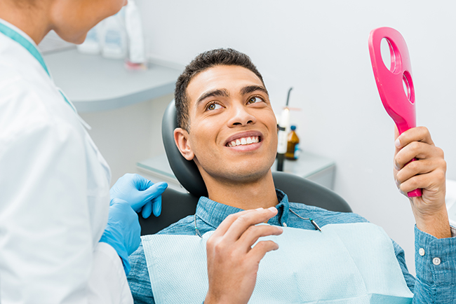
Regular oral exams are the backbone of predictable, long-lasting dental health. By combining careful clinical evaluation with targeted imaging and thoughtful preventative strategies, our team helps patients maintain healthy smiles and avoid more extensive treatment down the road. If you have questions about what to expect during an oral exam or whether you should schedule a visit, please contact us for more information.
An oral exam is a focused clinical assessment of your teeth, gums, bite, jaw function and the surrounding soft tissues. It allows the dental team to identify early signs of tooth decay, gum disease, wear patterns and other changes before they become more serious. Regular exams create a clear picture of your current oral health so care can be planned proactively.
Beyond spotting visible problems, an exam guides decisions about preventative care, home hygiene and any diagnostic imaging that may be needed. Early detection often means simpler, less invasive treatment and better long-term outcomes for your smile. Consistent exams also help establish a baseline so subtle changes can be tracked over time.
Your first visit begins with a review of medical and dental history so the team understands medications, past procedures and any health conditions that could affect care. The clinical exam includes inspection of each tooth, evaluation of gum tissues and assessment of your bite and jaw joint for signs of clenching, grinding or uneven forces. The provider also performs an oral soft-tissue exam and may discuss any symptoms you have noticed in the mouth, head or neck.
When indicated, diagnostic images such as bitewings or periapical films are taken to reveal areas not visible during the visual exam. After the assessment, the dentist or hygienist will summarize findings, answer your questions and outline a personalized plan for preventative care or treatment. This baseline visit sets the stage for regular monitoring and tailored recommendations going forward.
Oral health and whole-body health are closely connected, and an oral exam can reveal signs that relate to systemic conditions. Inflammation or infection in the mouth can contribute to broader health concerns, while conditions such as diabetes or certain medications often produce early changes inside the oral cavity. During an exam we look for clues like unexplained gum bleeding, persistent dry mouth or unusual lesions that may warrant medical coordination.
Identifying and managing oral disease reduces a potential source of chronic inflammation and supports overall wellness. When findings suggest a medical link, the dental team may recommend adjustments in scheduling, hygiene strategies or communication with your physician to help reduce risk and improve outcomes. This collaborative approach supports safe, effective care for both oral and general health.
The ideal interval for exams and professional cleanings depends on each patient’s risk profile, oral health history and current needs. Many patients benefit from twice-yearly visits, while people with higher risk for decay or periodontal disease may need more frequent monitoring. The dental team evaluates your individual factors to recommend an appropriate recall schedule that balances prevention with practical care.
Consistent preventative visits allow the hygienist to remove plaque and tartar in hard-to-reach areas and provide hands-on instruction for better daily hygiene. Regular exams also make it easier to detect early changes when treatments are typically less invasive and more predictable. Your recommended interval may change over time as your oral health or medical history evolves.
X-rays and three-dimensional imaging reveal conditions that cannot be seen during a visual exam, such as interproximal decay, root problems, bone loss or impacted teeth. Bitewing and periapical films are useful for evaluating crowns and roots, while panoramic images show broad jaw relationships and tooth development. In complex cases like implant planning or difficult extractions, CBCT provides detailed 3D anatomy that improves diagnostic accuracy and treatment safety.
Imaging is recommended selectively based on clinical findings and individual needs to ensure useful information is obtained while minimizing exposure. Digital radiography produces high-quality images quickly with reduced radiation compared with older film methods, and those images can be enhanced and stored for future comparison. The dentist will review any images with you and explain how they inform the recommended care.
Periapical films show an entire tooth from crown to root and are helpful for assessing root health, infections and nearby bone. Bitewing images focus on the crowns of molars and premolars and are especially useful for detecting early decay between teeth. Panoramic images provide a wide overview of both jaws and are valuable for evaluating tooth development, impactions and general jawbone form.
Cephalometric images are specialized views often used for orthodontic assessment, while CBCT captures volumetric data for three-dimensional evaluation of bone and anatomical structures. Each modality serves a distinct diagnostic purpose, and the dental team selects the right tool based on the clinical question at hand. When images are taken, they are reviewed with you so you understand what the pictures show and why certain options are recommended.
An oral cancer screening is an important part of a comprehensive exam and includes visual inspection and gentle palpation of the oral tissues, neck and jaw. The clinician looks for persistent sores, lumps, white or red patches, unexplained bleeding or any lesion that does not heal within a reasonable time. They will also review risk factors such as tobacco use, alcohol consumption and HPV exposure to inform the level of concern.
Early detection increases the likelihood of successful treatment, so any suspicious finding is documented and may prompt referral for biopsy or medical evaluation. The team explains what they observe, outlines next steps and coordinates care with specialists when appropriate. Regular screenings during routine exams are the most reliable way to spot early changes that deserve further attention.
While the components of an oral exam are similar, pediatric visits focus on growth, development and age-appropriate preventative strategies. For children we monitor tooth eruption, jaw development, spacing, oral habits and provide guidance on diet, home care and protective measures like sealants and mouthguards. Exams for young patients are also designed to build positive experiences and educate parents about long-term oral health habits.
Adult exams place additional emphasis on periodontal health, wear, restorative needs and conditions that emerge with age, such as increased risk of root decay or gum recession. The clinician adapts the exam and preventative recommendations to each life stage, medical history and risk profile. Whether for a child or an adult, the goal is early detection and a personalized plan that supports lasting oral health.
Prepare by bringing an up-to-date list of medications, a summary of relevant medical history and any questions or symptoms you want to discuss during the visit. Arrive having brushed your teeth so the clinician can see your oral surfaces clearly, but avoid heavy mouth rinses or chewing gum just before the appointment. If you wear removable appliances, bring them to the visit so they can be examined and evaluated for fit and hygiene.
If imaging is likely, leave jewelry and large accessories at home or remove them before the radiograph to avoid interference. For children, bring familiar comfort items and be ready to share information about diet, thumb-sucking or other habits that affect oral development. Clear communication about your concerns and history helps the dental team provide an efficient, targeted exam.
You should contact the office if you experience acute pain, noticeable swelling, persistent bleeding, a loose tooth or a sore that has not healed within two weeks. Sudden changes in bite, difficulty opening or closing the jaw, or any trauma to the mouth are also reasons to seek prompt evaluation. Early assessment of these issues can prevent complications and often leads to simpler, more effective treatment.
For non-urgent concerns or to discuss symptoms that are new or gradually worsening, reach out to schedule a timely appointment so the team can determine whether immediate care or an adjusted recall interval is appropriate. Clear communication helps ensure your comfort and protects long-term oral health, and the practice will guide you on the next steps based on the findings. If you are unsure whether a situation requires urgent attention, a quick phone call can help clarify the best course of action.
Email: